Dry AMD vs. Wet AMD: What's the Difference?
June 8, 2026
Age-related macular degeneration (AMD) is one of the leading causes of vision loss among older adults. It affects the macula—the central portion of the retina responsible for sharp, detailed vision—and can make everyday activities such as reading, driving, recognizing faces, and using a computer increasingly difficult. While AMD is generally divided into two main forms, dry AMD and wet AMD, understanding the difference between them is important because they behave differently and require different approaches to monitoring and treatment.
Dry AMD, also known as non-neovascular or nonexudative AMD, is by far the more common form. It develops when aging and other factors cause changes in the macula, including the accumulation of deposits called drusen beneath the retina. Small drusen can be a normal finding with aging, but larger and more numerous drusen can be associated with AMD. As dry AMD progresses, the retinal cells responsible for central vision can become damaged or lost.
For many people, dry AMD progresses slowly and may cause relatively mild vision changes for years. However, advanced dry AMD can lead to geographic atrophy (GA), in which areas of the retina gradually lose essential cells. Geographic atrophy can enlarge over time and cause permanent blind spots or significant loss of central vision. Importantly, advanced dry AMD is not simply a benign or inactive form of the disease—it can be a progressive and vision-threatening condition.
Wet AMD, also called neovascular AMD, occurs when abnormal blood vessels grow beneath or within the retina. These fragile vessels can leak fluid or blood, damaging the macula and causing vision loss. Wet AMD can develop rapidly, and the resulting vision changes may be much more sudden than those associated with typical dry AMD.
One of the reasons wet AMD requires prompt attention is that retinal damage can occur relatively quickly. Patients may notice new distortion of straight lines, a blurry or dark area in the center of their vision, decreased central vision, or difficulty reading. If these symptoms develop suddenly, an evaluation by an eye-care professional—ideally a retina specialist—should not be delayed.
The good news is that wet AMD is now highly treatable. The primary treatment involves anti-VEGF medications, which are injected directly into the eye. VEGF, or vascular endothelial growth factor, promotes the growth and leakage of abnormal blood vessels. By inhibiting VEGF, these medications can reduce leakage, control abnormal blood-vessel growth, and help preserve vision. In many patients, treatment can stabilize vision and, in some cases, improve it. Because wet AMD is typically a chronic condition, patients may require ongoing injections and monitoring.
Treatment for dry AMD depends on the stage and characteristics of the disease. For patients with intermediate AMD, specific vitamin and mineral formulations based on the AREDS2 study may be recommended in appropriate circumstances. However, these supplements are not appropriate for everyone and should be discussed with an eye-care professional.
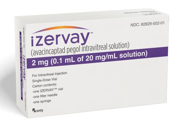
There have also been major advances in the treatment of geographic atrophy, the advanced form of dry AMD. FDA-approved complement inhibitors, including IZERVAY® (avacincaptad pegol) and SYFOVRE® (pegcetacoplan), are designed to slow the progression of geographic atrophy. These treatments do not restore retinal tissue that has already been lost, but they represent an important development in the management of advanced dry AMD.
Another important point is that dry AMD and wet AMD are not completely separate diseases. Wet AMD can develop in an eye that previously had dry AMD. For this reason, patients diagnosed with AMD should have regular retinal examinations and should monitor their vision for changes. Some patients may be advised to use an Amsler grid at home to help detect new distortion or changes in central vision.
Modern retinal imaging has also transformed the way AMD is diagnosed and monitored. Optical coherence tomography (OCT) allows a retina specialist to obtain high-resolution cross-sectional images of the retina and detect findings such as retinal fluid, pigment epithelial changes, drusen, and areas of atrophy. OCT and other imaging technologies allow physicians to identify subtle changes that may not yet be noticeable to the patient and to monitor the response to treatment over time.
The most important distinction to remember is that dry AMD is primarily associated with progressive retinal and macular cell loss, while wet AMD involves abnormal blood-vessel growth and leakage. Both forms can threaten central vision, but wet AMD can progress much more rapidly and requires prompt treatment when active disease is present.
If you have been diagnosed with AMD—or if you notice new distortion, flashes, floaters, blurred central vision, or a dark or missing area in your vision—it is important to have your eyes evaluated. Early detection, careful monitoring, and appropriate treatment can make a significant difference in preserving vision.
Advances in retinal imaging and treatment have changed the outlook for patients with AMD. While there is currently no cure for age-related macular degeneration, today's treatment options allow retina specialists to monitor the disease more precisely, treat active wet AMD effectively, and slow the progression of geographic atrophy in appropriate patients. Regular retinal care remains one of the most important steps you can take to protect your vision.